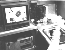
The planetary microscope (in gloved hand) and a TV image of lunar mineral and agglutinate particles.
Dr. Petr Jakés, longtime lunar investigator, has developed a miniature optical microscope for planetary field geology. Dr. Jakés brought his microscope to the Lunar Sample Laboratory, and demonstrated its capabilities for studying actual lunar soil.
In a recent presentation to the European Geophysical Society, Dr. Jakés asserted, "Identification of mineral phases, rock textures, particle shapes and sizes help interpret data on chemical composition of the studied/analyzed areas of a planetary surface and helps to understand the processes shaping it. Volcanic processes, impact, magmatic or aeolian processes, and mechanical or chemical weathering could be identified through the imaging of the surface if the resolution of the images is better than mineral grain size."
Dr. Jakés' microscope, about the size of a deck of cards, uses CCD chips and its own internal light source. It has a field of view of 3 mm and a resolution of approximately 10 µm. It produces a color image which can be viewed on a television screen, giving a magnification of around 100X. Using a digitizer and frame grabber, individual images can be saved to computer memory and manipulated with image analysis software. Filters can be placed in the light path to produce multispectral images.
For the lunar soil tests, samples that had been previously opened by other investigators and returned were selected. The tests were run in the PI experiment clean room in the Lunar Sample Laboratory. The authors, along with Linda Watts, prepared the samples for imaging and oversaw the tests. The Lunar and Planetary Institute provided the computer and frame grabber. Soil samples were poured onto a teflon disc and the microscope was simply placed over them.
Splits of mare soil 10084, which had previously been sieved and washed, provided excellent images. Particle size distributions were easily estimated. Characteristic features including rock and mineral fragments, agglutinates, and pyroclastic glass beads were immediately recognizable. Impact glass deposits around zap pits were especially prominent, due to the near-vertical illumination. The occasional exotic anorthite fragment was obvious.

The planetary microscope (in gloved hand) and a TV image of lunar mineral and agglutinate particles.
An untreated split of soil 14163 proved considerably
more challenging. This sample, like many lunar soils, included
around 10 wt% of particles smaller than 10µm. This fine material, at
or below the camera's resolution limit, coats every surface on
the larger particles. In our imaging tests the particles' sizes,
shapes, and colors were sometimes obscured. The best images
were obtained when the samples were first subjected to v
ibration, which caused the fine dust to segregate from the larger particles.
Dr. Jakés has taken a major step in support of the next generation of planetary robotic explorers. He has developed the space age version of the venerable geologist's hand lens. Now he has demonstrated this instrument's capabilities and limitations on actual lunar soil. One of the real challenges to be faced in humanity's next steps across the solar system is to make careful close-up observation of planetary surfaces. This experiment showed the value of testing new analytical equipment against real samples of extraterrestrial material.
The planetary microscope was developed at the Charles
University in Prague and the Astronomical Institute of the Czech
Academy, with additional support from Professor Wänke at the
Max Planck Institute. Dr. Jakés can be
contacted by email at jakes@prfdec.natur.cuni.cz.